
The standard paper speed is 25 mm/s. Hence one small square on the ECG is equivalent to 0.04 s; one large square is 0.2 s The quickest way to calculate the heart rate is to count the number of large squares between QRS complexes and divide into 300, e.g. if there are three large squares, the heart rate is 100 beats/min.
A heart rate of > 100 bpm is a tachycardia (Fig. 38); < 60 bpm is a bradycardia.
 |
| Sinus tachycardia |
 |
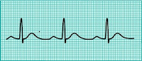
| Sinus bradycardia |
-Is it regular or irregular?
If there is any doubt, use a piece of paper to map out three or four consecutive
beats and see whether the rate is the same further along the ECG.
Regular rhythms
- P wave precedes every QRS complex with consistent PR interval is sinus rhythm.
- No discernable P wave preceding each QRS but narrow regular QRS complexes is a nodal or junctional rhythm.
- No discernable P waves preceding each QRS complex with an irregular rate is atrial fibrillation.
- P wave preceding each QRS with consistent PR interval, the rhythm is sinus arrhythmia.
- If P waves are present but there is progressive lengthening of the PR interval ending with non-conducted P wave (‘dropped beat’) followed by a normally conducted P wave with a shorter PR interval, the patient is in Wenckebach’s (or Mobitz type I) 2nd degree AV block.
There is nothing mysterious about working out the cardiac (or QRS) axis. It represents
the net depolarization through the myocardium and is worked out using the
limb leads, in particular leads I and aVF. The directions of each of these leads
(the cardiac vector) are summarized in Fig. 40. By convention, the direction of lead
I is 0 degree; and aVF points down (aV‘FEET’).
The rules for working out the cardiac axis are as follows:
- Calculate the net deflection of each lead – e.g. in lead I, if there is a Q wave measuring three small squares and an R wave height of six small squares, the net deflection is þ3. Do this for leads I and aVF.
- A net positive deflection goes in the direction of the vector; negative deflections go in the opposite direction of the vector – e.g. net deflection of þ3 in I goes 3 points in the direction of I; a net deflection of -5 in aVF goes in the opposite deflection of the vector (i.e. upwards) by 5 points.
- The cardiac vector is therefore the sum of the individual vectors from I and aVF – e.g. +3 in I, -5 in aVF gives a vector of about 60 degree. A normal axis is between 0 degree and 90 degree; anything to the left of 0degree is termed left axis deviation; anything to the right of 90 degree is right axis deviation
Look at the P wave shape.
-Peaked P waves (P pulmonale) suggest right atrial hypertrophy – e.g. pulmonary
hypertension or tricuspid stenosis.
 |
| Tall P wave |
 |
| Bifid P wave |
PR interval
The PR interval is measured from the beginning of the P wave to the R wave and is usually 1 large square in duration (0.2 s). A short PR interval represents rapid conduction across the AV node, usually through an accessory pathway (e.g. Wolff–Parkinson–White syndrome)
 |
| Short PR interval |
 |
| First degree AV block |
A PR interval that lengthens with each consecutive QRS complex, followed by a P wave which has no QRS complex and then by a P wave with a short PR interval, is Wenckebach’s (or Mobitz type I) 2nd degree AV block
 |
| Second degree AV block; Mobitz I |
 |
| Second degree AV block; Mobitz II |
If the P waves are regular (usually at a rate of about 90) and the QRS complexes are regular (heart rate about 40 bpm), but there is no association between the two, then the rhythm is complete (or 3rd degree) AV block. This rhythm will need to be discussed with your seniors as will usually require cardiac pacing, and if the patient is compromised, e.g. hypotensive, will need insertion of a temporary pacing wire.
 |
| Third degree AV block |
First, look at the width of the QRS, then the morphology.
Normal QRS duration is less than three small squares (0.12 s) and represents normal conduction through the AV node and the bundle of His.
A broad QRS complex signifies either:
1. The beat is ventricular in origin, e.g. an ectopic beat, or
2. There is a bundle branch block.
A broad QRS complex with an RSR pattern in V1 represents right bundle branch block.
A broad QRS with an ‘M’ pattern in lead I represents left bundle branch block
.
 |
| Wide QRS (showing LBBB in lead 1) |
 |
| Deep Q wave |
ST segments
There are basically three abnormalities seen in the ST segement:
 |
| 1. ST depression – could signify cardiac ischaemia |
 |
| 2. ST elevation – highly suggestive of infarction |
3. ‘Saddle shaped’ – concave ST segments usually seen across all the ECG, suggesting a diagnosis of pericarditis.
If there is any evidence of ST segment abnormality, particularly in the context of a patient with chest pain, seek senior advice at once. It is important to note that ST segments are abnormal and cannot be interpreted
in patients with bundle branch block, especially LBBB.
QT interval
The QT interval is usually about 0.4 s (two large squares) and is important as prolongation can lead to serious ventricular arrhythmias such as torsades de pointes. It can be prolonged for several reasons – including drugs such as amiodarone, sotalol and some anti-histamines – so a drug history is crucial if this abnormality is seen. A family history of sudden cardiac death is also important as a congenital long QT syndrome may be present.
T waves
T waves should be upright in all leads other than leads III and V1 where an inverted
T wave can be a normal variant.
Tall tented T waves could represent hyperkalaemia.
 |
| Tall T wave |
 |
| T wave inversion |
T wave inversion can represent coronary ischaemia, previous infarction or electrolyte
abnormality such as hypokalaemia
Arrhythmias
Arrhythmias – be they tachyarrhythmias or bradyarrhythmias – should be treated in the context of the patient’s clinical condition. Thus, vital signs – e.g. blood pressure, oxygen saturations, conscious level – should all be known before treatment is instituted. You are much more concerned about the patient with a heart rate of 140 bpm and a systolic blood pressure of 60 mmHg than you are about the patient with a heart rate of 40 bpm with a normal blood pressure eating their lunch.
Bradyarrhythmias
Any heart rate below 60 bpm is considered a bradycardia. This heart rhythm could be anything from a sinus bradycardia or atrial fibrillation with a low ventricular rate to some form of heart block as outlined above.
Treatment of a compromised patient usually requires the insertion of a temporary pacing wire and your seniors should be alerted immediately in the event.
Tachyarrhythmias
Patients with tachyarrhythmias and any evidence of haemodynamic compromise should be considered for emergency DC cardioversion.
There are broadly two types of tachyarrythmia:
1. Narrow complex (QRS duration <120 ms)
2. Broad complex (QRS duration >120 ms)
Narrow complex tachycardias are supra-ventricular (i.e. above the ventricle) in origin. If the rhythm is irregular, the likely diagnosis is that of atrial fibrillation with a fast ventricular response.
 |
| Atrial fibrillation |
 |
| Atrial flutter with 2:1 block |
 |
| AV re-entrant tachycardia |
- ventricular in origin (ventricular tachycardia (VT)) or
- supra-ventricular in origin but have a pre-existing or rate-related bundle branch block. An old ECG may be helpful in this group.
The following are useful features to look out for that favour a diagnosis of VT over
an SVT with bundle branch block:
- capture beats – normal narrow complex beats ‘captured’ between the broad beats of the tachycardia.
 |
| Ventricular tachycardia with capture beat |
- fusion beats – similar to capture beats but the narrow QRS complex is superimposed by the broad ventricular beats.
- extreme axis deviation, usually left axis deviation.
- chest lead concordance – the chest leads V1–V6 all point the same way, i.e. all positive deflections or all predominantly negative directions.


