
Showing posts with label Pathoyphysiology. Show all posts
Showing posts with label Pathoyphysiology. Show all posts
Friday, November 30, 2012
Pathophysiology of Dengue Hemorrhagic Fever


Pathophysiology of Chronic Kidney Failure Secondary to DM Type II

The underlying pathophysiology defect in type 2 diabetes is characterized by the following three disorders (1) peripheral resistance to insulin, especially in muscles cells: (2) increased production of glucose by the liver, and (3) altered pancreatic secretion. Increased tissue resistance to insulin generally occurs first and eventually followed by impaired insulin secretions. The pancreas produces insulin, yet insulin resistance prevents its proper use at the cellular level. Glucose cannot enter target cells and accumulates in the blood streams, resulting in hyperglycemia. The high blood glucose levels often stimulate an increase in insulin production by the pancreas: thus. Type 2 diabetic individuals often have excessive insulin production (hyperinsulinemia).
Insulin resistance refers to tissue sensitivity to insulin. Intracellular reaction are diminished, making insulin less effective at stimulating glucose uptake by the tissues and regulating glucose release by the liver.
If blood glucose levels are elevated consistently for a significant period of time, the kidney’s filtration mechanism is stressed, allowing blood proteins to leak into the urine. As a result, the pressure in the blood vessels of the kidney increases. It is thought that the elevated pressure serves as the stimulus the level of nephropathy.
The earliest detectable change in the course of diabetic nephropathy is a thickening in the glomerulus. At this stage, the kidney may start allowing more albumin (protein) than normal in the urine, and this can be detected by sensitive tests for albumin. As diabetic nephropathy progresses, increasing numbers of glomeruli are destroyed. Now the amounts of albumin being excreted in the urine increases, and may be detected by ordinary urinalysis techniques. At this stage, a kidney biopsy clearly shows diabetic nephropathy and eventually leads to Chronic renal failure.
Sunday, April 29, 2012
Pathophysiology of Hypovolemic Shock
Tuesday, April 17, 2012
Pathophysiology of Pneumonia

Understanding pathophysiology of pneumonia will help you to point out the symptoms of pneumonia before they become fatal. a person suffering from pneumonia will usually experience fever chills and they may seem as if they are having difficulty during breathing. Most people will also complain of chest pains which could be accompanied by cough that has green or yellowish mucus. This is a fatal condition and it affects mostly the old and children who have a weak immunity.
Pathophysiology of pneumonia will assist you in knowing what the different causes of pneumonia are. They include lung injuries although this is no so common and only happens when someone has been involved in an accident. Flea bites are also said to be a cause of pneumonia but usually this is not very common. The major cause and which many of us know is the fungal, bacteria or viral infection of the respiratory tract which causes the fluid or air sac to form within the lungs.
Generally the pathophysiology of pneumonia is complicated and a thorough understanding of what causes the disease is very helpful. Pathophysiology of pneumonia will help you understand how to deal with the patient and the medication to be administered when an emergency occurs. There are different levels of pneumonia and in all cases special care is given to the patient to prevent fatalities.
Pathophysiology of Chronic Bronchitis

Pathophysiology of Thrombophlebitis

Pathophysiology of Emphysema
The pathophysiology of emphysema is best explained on the basis of decreased pulmonary elastic recoil. At any pleural pressure, the lung volume is higher than normal. Additionally, the altered relation between pleural and alveolar pressure facilitates expiratory dynamic compression of airways. Such compression limits airflow during forced expiration and, in severe instances, during tidal expiration. Another factor contributing to airflow limitation is disease of the airways, both large and small. In general, patients with relatively pure emphysema maintain blood gases in or near the normal range until very late in their course. PaO2 is maintained because of the preserved matching of ventilation and perfusion as alveolar walls are destroyed. PaCO2 is maintained because the ventilatory response to CO2 is not usually impaired. It is not clear why patients who are categorized clinically as "chronic bronchitics" are more likely to respond to an increased flow-resistive work of breathing by hypoventilating. Physical findings in emphysema are not specific. Radiologic changes are insensitive and are of less value than physiologic measurements.
Tuesday, September 7, 2010
Saturday, September 4, 2010
Pathophysiology of Diabetes Mellitus

The pathophysiology of diabetes mellitus (all types) is related to the hormone insulin, which is secreted by the beta cells of the pancreas. This hormone is responsible for maintaining glucose level in the blood. It allows the body cells to use glucose as a main energy source. However, in a diabetic person, due to abnormal insulin metabolism, the body cells and tissues do not make use of glucose from the blood, resulting in an elevated level of blood glucose or hyperglycemia. Over a period of time, high glucose level in the bloodstream can lead to severe complications, such as eye disorders, cardiovascular diseases, kidney damage and nerve problems.
Tuesday, August 24, 2010
Pathophysiology of Schizophrenia

The pathophysiology of schizophrenia has long remained a mystery and still today, even with various hypotheses, remains somewhat uncertain: there are too many variants; not enough consistency in findings; and, despite research, a lack of documented proof.
Dopamine is both a hormone and a neurotransmitter, which means that it activates five different receptors in the brain, aptly named D1, D2, D3, D4, and D5. That said, it may not be the only neurotransmitter involved in the pathophysiology of schizophrenia. Glutamate and Serotonin have also been implicated. .
Contributing to this hypothesis is the fact that drugs administered to aid dopaminergic activity bring on schizophrenic characteristics such as psychosis, in a patient, whereas drugs administered to block them help reduce, or eliminate symptoms of schizophrenia altogether.
Additional studies affecting the pathophysiology of schizophrenia include suggestions that maternal factors such as infection, malnutrician, location of birth, season of birth, and delivery, may play a significant part in the formation and subsequent appearance of schizophrenia. Studies have shown that the worldwide rate of births affected with schizophrenia is up to 8% higher when occurring in spring or winter, though no explanation for this can be offered.

Another aspect of the pathophysiology of schizophrenia that has been explored in relative detail is that of genetics, and their relation to the likelihood of immediate relatives being born with the disease. Shockingly, it has been found that 10% of all immediate family members of an infected person will be struck down with the disease. This is specifically in relation to parents, siblings, and children. With regards to twins or other multiple births, the chances they will share the disease is 50%. Genetic reports suggest that it is the X chromosome which determines whether or not a person is infected with schizophrenia, specifically, chromosomes 1, 3, 5, and 11, however further studies are needed in order to prove this theory.
Though there are many theories and hypotheses regarding the pathophysiology of schizophrenia, there is, unfortunately, still no cure for the disease. The best a sufferer can hope for nowadays is to benefit from available medication which keeps the disease under control or in remission for the duration of time for which it is taken.
Pathophysiology of Bronchial Asthma

1. An asthma attack may occur spontaneously or in response to a trigger. Either way, the attack progresses in the following manner:
- There is an initial release of inflammatory mediators from bronchial mast cells, epithelial cells, and macrophages, followed by activation of other inflammatory cells
- Alteration of autonomic neural control of airway tone and epithelial integrity occur and the increased responsiveness in airways smooth muscle results in clinical manifestations (e.g. wheezing and dyspnea)
- Bronchial spasm
- Inflammation and edema of the mucosa
- Production of thick mucus, which results in increased airway resistance, premature closure of airways, hyperinflation, increased work of breathing, and impaired gas exchange
Bronchial Asthma - Nursing Care Plan
Pathophysiology of Amoebiasis
Mature cyst in the large intestines leaves the host in great numbers (the host remains asymptomatic). The cyst can remain viable and infective in moist and cool environment for at least 12 days, and in water for 30 days. The cysts are resistant to levels of chlorine normally used for water purification. They are rapidly killed by purification, desiccation and temperatures below 5 and above 40 degrees.

The metacystic trophozoites of their progenies reach the cecum and those that come in contact with the oral mucosa penetrate or invade the epithelium by lytic digestion.
The trophozoites burrow deeper with tendency to spread laterally or continue the lysis of cells until they reach the sub-mucosa forming flash-shape ulcers. There may be several points of penetration.
From the primary site of invasion, secondary lesions maybe produced at the lower level of the large intestine.
Progenies of the initial colonies are squeezed out to the lower portion of the bowel and thus, have the opportunity to invade and produce additional ulcers. Eventually, the whole colon may be involved.
E. histolytica has been demonstrated in practically every soft organ of the body.
Trophozoites which reach the muscularis mucosa frequently erode the lymphatics or walls of the mesenteric venules in the floor of the ulcers, and are carried to the intrahepatic portal vein.
If thrombi occur in the small branches of the portal veins, the trophozoites in thrombi cause lytic necrosis on the wall of the vessels and digest a pathway into the lobules.
The colonies increase in size and develop into abscess.
A typical liver abscess develops and consists of:
- Central zone necrosis
- Median zone of stoma only
- An outer zone of normal tissue newly invaded by amoeba. Most amoebic abscess of the liver are in the right lobe.
Source:
Handbook of Common Communicable and Infectious Diseases 2006 Edition by Dionesia Mondejar-Navales, RN, MAEd
Pathophysiology of Liver Cirrhosis

Cirrhosis is known in three major forms. In Laennec’s (alcohol-induced) cirrhosis, fibrosis occurs mainly around central veins and portal areas. This is the most common form of cirrhosis and results from chronic alcoholism and malnutrition. Postnecrotic (micronodular) cirrhosis consist of broad bands of scar tissue and results from previous acute viral hepatitis or drug-induced massive hepatic necrosis. Biliary cirrhosis consists of scarring of bile ducts and lobes of the liver and results from chronic biliary obstruction and infection (cholangitis), and is much rarer than the preceding forms.
Pathophysiology of Congestive Heart Failure (CHF)

Heart Failure, also known as Congestive Heart Failure, is a clinical syndrome that results from the progressive process of remodeling, in which mechanical and biochemical forces alter the size, shape, and function of the ventricle’s ability to pump enough oxygenated blood to meet the body’s metabolic requirements. Compensatory mechanisms of increased heart rate, vasoconstriction, and hypertrophy eventually fail, leading to the characteristic syndrome of heart failure: Elevated ventricular or atrial pressures, sodium and water retention, decreased cardiac output, and circulatory and pulmonary congestion. Systolic dysfunction occurs when the left ventricle is unable to relax and fill sufficiently to accommodate enough oxygenated blood returning from the pulmonary circuit. Systolic dysfunction leads to increased vascular resistance and increased afterload. Diastolic dysfunction leads to pulmonary vascular congestion.
Pathophysiology of Hypertension

The two major types of hypertension are primary (essential) hypertension, in which diastrolic pressure is 90 mm Hg or higher and systolic pressure is 140 mm Hg or higher in absence of other causes of hypertension (approximately 95 % of patients); and Secondary hypertension, which results primarily from renal disease, endocrine disorders, and coarctation of the aorta. Either of these conditions may give rise to accelerated hypertension – a medical emergency – in which blood pressure elevates very rapidly to threaten one or more of the target organs: the brain, kidney, or the heart.
Hypertension is one of the most prevalent chronic diseases for which treatment is available; however, most patients with hypertension are unaware, untreated, or inadequately treated. Risk factors for hypertension are age between 30 and 70; black; overweight; sleep apnea; family history; cigarette smoking; sedentary lifestyle; and diabetes mellitus. Because hypertension presents no over symptoms, it is termed the “silent killer.” The untreated disease may progress to retinopathy, renal failure, coronary artery disease, heart failure, and stroke.
Hypertension in children is defined as the average systolic or diastolic blood pressure greater than or equal to the 95th percentile for age and sex with measurement on at lease three occasions. The incidence of hypertension in children is low, but it is increasingly being recognized in adolescents; and it may occur in neonates, infants, and young children with secondary causes.
Pathophysiology of Cholelithiasis/Cholecystitis

Most gallstones result from supersaturation of cholesterol in the bile, which acts as an irritant, producing inflammation in the gallbladder, and which precipitates out of bile, causing stones. Risk factors include gender (women four times as like to develop cholesterol stones as men), age (older than age 40), multiple parity, obesity, use of estrogen and cholesterol-lowering drugs, bile acid malabsorption with GI disease, genetic predisposition, rapid weight loss. Pigment stones occur when free bilirubin combines with calcium. These stones occur primarily in patients with cirrhosis, hemolysis, and biliary infections.
Acute cholecystitis is caused primarily by gallstone obstruction of the cystic duct with edema, inflammation, and bacterial invasion. It may also occur in the absence of stones, as a result of major surgical procedures, severe trauma, or burns.
Chronic cholecystitis results from repeated attacks of cholecystitis, presence of stones, or chronic irritation. The gallbladder becomes thickened, rigid, fibrotic, and functions poorly.
Complications of gallbladder disease include cholangitis; necrosis, empyema, and perforation of gallbladder; biliary fistula through duodenum; gallstone ileus; and adenocarcinoma of the gallbladder.
Pathophysiology of Leukemia

Although the cause of leukemias is unknown, predisposing factors include genetic susceptibility, exposure to ionizing radiation or certain chemicals and toxins, some genetic disorder (Down syndromes, Fanconi’s anemia), and human T-cell leukemia-lymphoma virus. Complications include infection, leukostasis leading to hemorrhage, renal failure, tumor lysis syndrome, and disseminating intravascular coagulation.
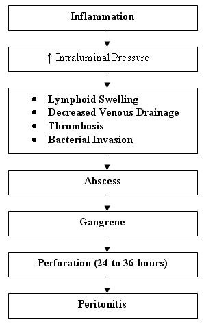
Pathophysiology of Appendicitis

Saturday, August 21, 2010
Pathophysiology of Chronic Obstructive Pulmonary Disease (COPD)

Chronic bronchitis is chronic inflammation of the lower airways characterized by excessive secretion of mucus, hypertrophy of mucous glands, and recurring infection, progressing to narrowing and obstruction of airflow. Emphysema is the enlargement of the air spaces distal to the terminal bronchioles, with breakdown of alveolar walls and loss of elastic recoil of the lungs. The two conditions may overlap, resulting in subsequent derangement of airways dynamics (e.g., obstruction to airflow). In pulmonary emphysema, lung function progressively deteriorates for many years before the illness becomes apparent.
The most common cause of COPD is cigarette smoking. Air pollution, occupational exposures, allergens, and infections may also act as irritants. Alpha1-antitrypsin deficient is an infrequent cause. Complications include respiratory failure, pneumonia or other overwhelming respiratory infection, right heart failure (cor pulmonale), arrhythmias, and depression.


Subscribe to:
Posts (Atom)